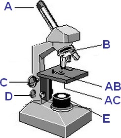
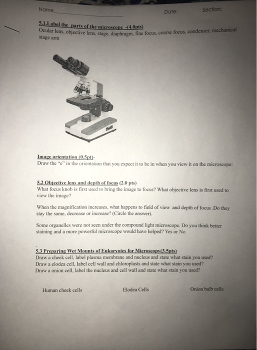
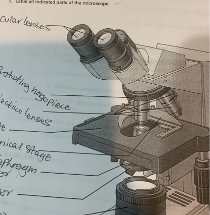
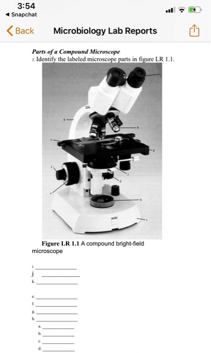
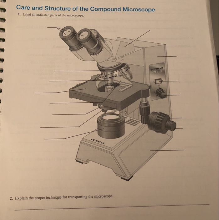
43 label all indicated parts of the microscope
Answered: 5) Label the parts of the microscope. 1… | bartleby Name the part of the microscope that you use to control the cone of light coming through the… A: An instrument which functions by processing the light waves to enhance the image that is to be… A: The microscope is the major tool in the identification of a microorganism after its isolation. Compound Microscope: Parts of Compound Microscope - BYJUS The parts of the compound microscope can be categorized into: Mechanical parts; Optical parts (A) Mechanical Parts of a Compound Microscope. 1. Foot or base. It is a U-shaped structure and supports the entire weight of the compound microscope. 2. Pillar. It is a vertical projection. This stands by resting on the base and supports the stage. 3. Arm
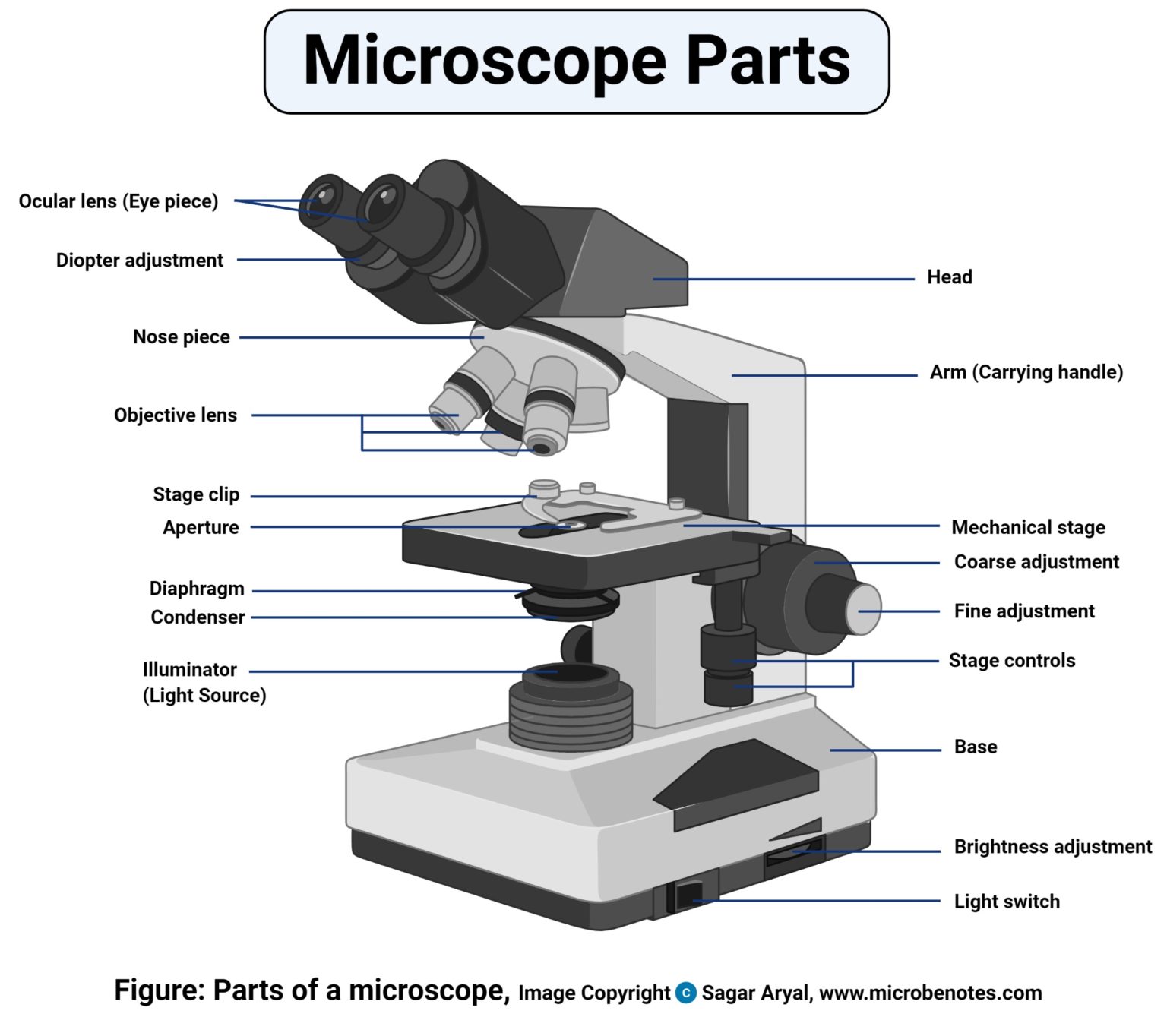
Parts of a microscope with functions and labeled diagram Apr 19, 2022 · These parts include: Eyepiece – also known as the ocular. This is the part used to look through the microscope. Its found at the top of the... Eyepiece tube – it’s the eyepiece holder. It carries the eyepiece just above the objective lens. In some microscopes... Objective lenses – These are the ...
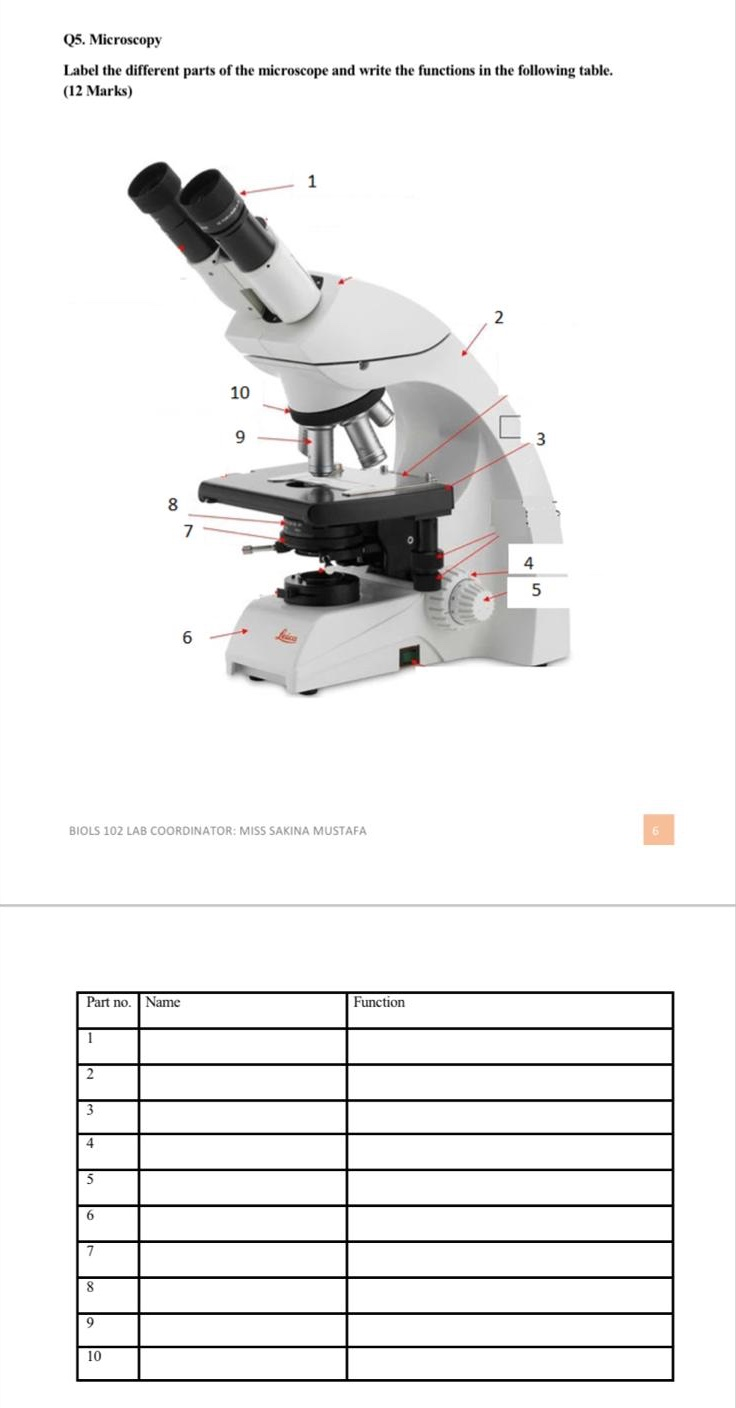
Label all indicated parts of the microscope
Using the Microscope: Basic Tutorial: Part 2: Components. - Micrographia The Eye. The cornea and the eye lens are the final optical components in the image-forming path to the retina. In a person with normal vision, the eyelens will be relaxed as though the eye is forming an image of a very distant object, and the focusing controls on the microscope used to achieve image sharpness. Microscope Quiz: How Much You Know About Microscope Parts ... - ProProfs Projects light upwards through the diaphragm, the specimen, and the lenses. 5. Is used to regulates the amount of light on the specimen. Supports the slide being viewed. Moves the stage up and down for focusing. 6. Is used to support the microscope when carried. Moves the stage slightly to sharpen the image. Parts of the Microscope with Labeling (also Free Printouts) A microscope is one of the invaluable tools in the laboratory setting. It is used to observe things that cannot be seen by the naked eye. Table of Contents 1. Eyepiece 2. Body tube/Head 3. Turret/Nose piece 4. Objective lenses 5. Knobs (fine and coarse) 6. Stage and stage clips 7. Aperture 9. Condenser 10. Condenser focus knob 11. Iris diaphragm
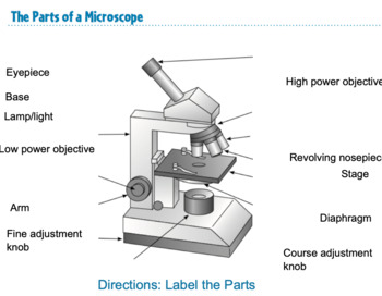
Label all indicated parts of the microscope. Activity 3.1.docx - Denise Learned Bio201 The Microscope... Denise Learned Bio201 The Microscope Care and Structure of the Compound Microscope 1. Label all indicated parts of the microscope. 2. Explain the proper technique for transporting the microscope. Answer: HOLD IT IN AN UPRIGHT POSITION WITH ONE HAND ON ITS ARM AND THE OTHER SUPPORTING ITS BASE A Study of the Microscope and its Functions With a Labeled Diagram ... These labeled microscope diagrams and the functions of its various parts, attempt to simplify the microscope for you. However, as the saying goes, 'practice makes perfect', here is a blank compound microscope diagram and blank electron microscope diagram to label. Download the diagrams and practice labeling the different parts of these ... Parts of a Microscope - Lab - Perkins School for the Blind Braille or large print handout including microscope parts and their functions. 1. Make braille or large print labels for the microscope. These should be letters, a through j. 2. Label the parts of the microscope with either large print or braille letters (as per the picture). 3. Label parts on microscope Flashcards | Quizlet Created by ashleibryson Terms in this set (14) Eyepiece Body Tube on microscope High power objective Low power objective Stage Opening Diaphragm Lever Light on microscope Coarse adjustment Fine adjustment Nosepiece Arm on a microscope Stage Clip Stage on microscope Base Sets found in the same folder Cell Organelles 28 terms MKL67
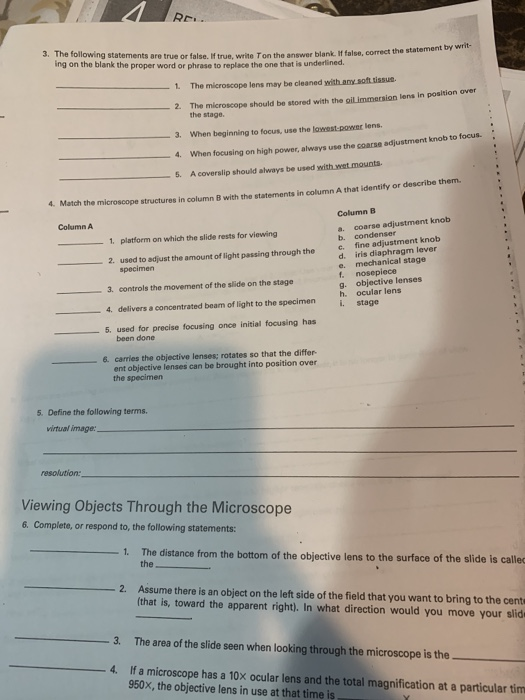
Compound Microscope Parts The three basic, structural components of a compound microscope are the head, base and arm. Head/Body houses the optical parts in the upper part of the microscope. Base of the microscope supports the microscope and houses the illuminator. Arm connects to the base and supports the microscope head. It is also used to carry the microscope. openlab.citytech.cuny.edu The Microscope EXERCSE Mar; qldb Lab Time/Date Name Care and Structure of the Compound Microscope I. Label all indicated parts of the microscope. esQi€ce 2. Explain the proper technique for transporting the microscope. 0b qs Ont en arm loose. 33 . 34 Review Sheet 3 3. Each of the following statements is either true or false. PDF Microscope Parts and Functions - WPMU DEV Microscope Parts and Functions Microscope One or more lenses that makes an enlarged image of an object. 8/7/2018 2 •Simple •Compound •Stereoscopic ... Always carry a microscope with one hand holding the arm and one hand under the base. Base Diaphragm A B Carrying a Microscope. 8/7/2018 13 Compound Microscope Parts - Labeled Diagram and their Functions There are three major structural parts of a compound microscope. The head includes the upper part of the microscope, which houses the most critical optical components, and the eyepiece tube of the microscope. The base acts as the foundation of microscopes and houses the illuminator. The arm connects between the base and the head parts.
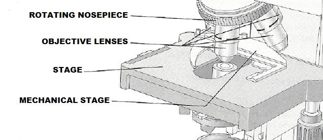
PDF The Microscope - Holly H. Nash-Rule, PhD After the parts of the microscope have been identified, turn on the light and adjust the interpupillary distance so that ... Care and Structure of the Compound Microscope 1. Label all indicated parts of the microscope. Ocular lenses Rotating nosepiece Objective lenses Stage Mechanical stage Iris diaphragm lever Condenser Substage light Head Arm (Solved) - Care and Structure of the Compound Microscope 1. Label all ... Care and Structure of the ... Label the microscope — Science Learning Hub All microscopes share features in common. In this interactive, you can label the different parts of a microscope. Use this with the Microscope parts activity to help students identify and label the main parts of a microscope and then describe their functions. Drag and drop the text labels onto the microscope diagram. Solved > 1. Label all indicated parts of the microscope. from Chapter 3 ... Solved expert answers for Human Anatomy and Physiology Laboratory Manual, Main Version 10th Edition by Elaine N. Marieb, Susan J. Mitchell, Lori A. Smith. Instant access with 24/7 expert assistance.
Labeling the Parts of the Microscope | Microscope World Resources Labeling the Parts of the Microscope. This activity has been designed for use in homes and schools. Each microscope layout (both blank and the version with answers) are available as PDF downloads. You can view a more in-depth review of each part of the microscope here. Download the Label the Parts of the Microscope PDF printable version here.
Microscope Parts & Functions - AmScope Body: Often referred to as the head, the body is the upper part of a microscope including, eyepieces and objectives. Most modern microscopes are modular in the sense that the same body can be used with different bases and vice versa.
MasteringMicrobiology - Week 1 Post Lab Flashcards | Quizlet Label parts of the microscope. (From top to bottom) Objective Lens Slide Clip Stage Label the indicated parts of the microscope. (From top to bottom and left to right) Ocular Lens Objective Lens Oil Immersion Lens Navigation Knobs Coarse Focus Fine Focus Which of the following describes streptococci? Chains of spherically shaped cells
Solved Care and Structure of the Compound Microscope 1. - Chegg Anatomy and Physiology Anatomy and Physiology questions and answers Care and Structure of the Compound Microscope 1. Label all indicated parts of the microscope. Cvlar len hy 3. The following statements are true or false. If true, write ing on the blank the proper word or phrase to replace the one that is underlined. Ton the answer blank.
Molecular Expressions Microscopy Primer: Anatomy of the Microscope ... Color Codes - Microscope manufacturers label their objectives with color codes to help in rapid identification of the magnification and any specialized immersion media requirements. The dark blue color code on the objective illustrated in Figure 1 indicates the linear magnification is 60x.
Microscope Parts and Functions The microscope parts work together in hospitals and in forensic labs, for scientists and students, bacteriologists and biologists so that they may view bacteria, plant and animal cells and tissues, and various microorganisms the world over.
Exercise 3: The Microscope Flashcards - Easy Notecards LABEL ALL INDICATED PARTS OF THE MICROSCOPE. 6 EXPLAIN THE PROPER TECHNIQUE FOR TRANSPORTING THE MICROSCOPE. WHEN TRANSPORTING THE MICROSCOPE, HOLD IT IN AN UPRIGHT POSITION WITH ONE HAND ON ITS ARM AND THE OTHER SUPPORTING ITS BASE. AVOID SWINGING THE INSTRUMENT DURING ITS TRANSPORT AND JARRING THE INSTRUMENT WHEN SETTING IT DOWN. 7
Microscope, Microscope Parts, Labeled Diagram, and Functions The description given below summarize the brief description of microscope parts used to visualize the microscopic specimens such as animal cells, plant cells, microbes, bacteria, viruses, microorganisms etc. The Microscopes parts divided into three different structural parts Head, Base, and Arms.
LABEL ALL INDICATED PARTS OF THE MICROSCOPE.docx - Course Hero LABEL ALL INDICATED PARTS OF THE MICROSCOPE. 2. LABEL ALL INDICATED PARTS OF THE MICROSCOPE. 3. LABEL ALL INDICATED PARTS OF THE MICROSCOPE. 4 LABEL ALL INDICATED PARTS OF THE MICROSCOPE. 5 LABEL ALL INDICATED PARTS OF THE MICROSCOPE. 6 EXPLAIN THE PROPER TECHNIQUE FOR TRANSPORTING THE MICROSCOPE. WHEN TRANSPORTING THE MICROSCOPE, HOLD IT IN AN UPRIGHT POSITION WITH ONE HAND ON ITS ARM AND THE OTHER SUPPORTING ITS BASE.
Solved Care and Structure of the Compound Microscope 1. - Chegg Expert Answer 100% (1 rating) Answer 1) A. Ocular lens. B. Rotating nose piece. C. Stage. D. Condensor. E. Iris diaphragm. F. Light source. G. Base. H. Arm. I. Objective lens. J. Mechanical stage. K. Condensor knob. L. Coarse adjustment. M. Fine adjustment. N. Light control. Micr … View the full answer
City University of New York Label all indicated parts of the microscope. Occolar (OSU uuhamca| clenor In's 2. Explain the proper technique for transporting the microscope. sj+s onQ flan/ 10 arqQ odd lcnÒb Bose OLYMPUS . 36 Review Sheet 3 3. The following statements are true or false. If true, write Ton the answer blank. If false, correct the statement by
Parts of the Microscope with Labeling (also Free Printouts) A microscope is one of the invaluable tools in the laboratory setting. It is used to observe things that cannot be seen by the naked eye. Table of Contents 1. Eyepiece 2. Body tube/Head 3. Turret/Nose piece 4. Objective lenses 5. Knobs (fine and coarse) 6. Stage and stage clips 7. Aperture 9. Condenser 10. Condenser focus knob 11. Iris diaphragm
Microscope Quiz: How Much You Know About Microscope Parts ... - ProProfs Projects light upwards through the diaphragm, the specimen, and the lenses. 5. Is used to regulates the amount of light on the specimen. Supports the slide being viewed. Moves the stage up and down for focusing. 6. Is used to support the microscope when carried. Moves the stage slightly to sharpen the image.
Using the Microscope: Basic Tutorial: Part 2: Components. - Micrographia The Eye. The cornea and the eye lens are the final optical components in the image-forming path to the retina. In a person with normal vision, the eyelens will be relaxed as though the eye is forming an image of a very distant object, and the focusing controls on the microscope used to achieve image sharpness.
















Post a Comment for "43 label all indicated parts of the microscope"