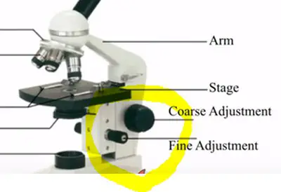
42 label the image of a compound light microscope using the terms provided
How to Use a Compound Microscope: 11 Steps (with Pictures) - wikiHow Focus the microscope. Looking through the eyepiece, arrange the illuminator and the diaphragm to reach the most comfortable level of light. Move the specimen slide so that the image is in the center of your view. [10] Arrange the illuminator until you've arrived at a comfortable level of light. Parts of the Microscope Printables - ThoughtCo Most microscopes used in a classroom setting are compound microscopes. These usually consist of a light source and three to five lenses with a total magnification of 40x to 1000x. The following free printables can help you teach your students the basic parts of a microscope so that they are ready to dive into a world previously unseen.
Label Parts Of A Compound Microscope Teaching Resources | TpT Microscope Quiz by Quick Witted Owl Lesson Plans Made Easy 3 $1.99 PDF Students will need to label the parts of a compound light microscope and answer a 8 easy questions. A word bank is given at the bottom of the quiz.

Label the image of a compound light microscope using the terms provided
Virtual Lab 2 Microscopy and Cells_for Hybrid absense.docx... return to the bionetwork's virtual microscope: o - tools/virtual-microscope o from the main menu, click "explore". o in the next screen, click on the question mark that is over the box of microscope slides. o from the slide catalog menu, select "sample slides" and then "letter e". o you now have to use the proper steps to bring the specimen into … Labeling the Parts of the Microscope Labeling the Parts of the Microscope This activity has been designed for use in homes and schools. Each microscope layout (both blank and the version with answers) are available as PDF downloads. You can view a more in-depth review of each part of the microscope here. Download the Label the Parts of the Microscope PDF printable version here. Solved Microscope parts/labeling 9 Label the image of a Question: Microscope parts/labeling 9 Label the image of a compound light microscope using the terms provided. 1 points eyepiece eyepiece light source ...
Label the image of a compound light microscope using the terms provided. Label the image of a compound light microscope using the ... Is this correct?? Label the image of a compound light microscope using the terms provided. compound microscope parts (labeling) Flashcards - Quizlet Start studying compound microscope parts (labeling). Learn vocabulary, terms, and more with flashcards, games, and other study tools. ... light source of the microscope. what is 8? eyepiece (ocular lens) - magnifying piece that is looked into in order to see the specimen ... knob that brings the image to a sharper focus. what is 13? base - the ... Compound Light Microscope Flashcards | Quizlet Start studying Compound Light Microscope. Learn vocabulary, terms, and more with flashcards, games, and other study tools. Home. ... -Use an electron beam to create magnified images of objects-Primarily found in research and human medical facilities. Compound light microscope ... 132 terms. quizlette835084 PLUS. Blood Components. 45 terms. hope ... Light Microscope- Definition, Principle, Types, Parts, Labeled Diagram ... The difference is simple light microscopes use a single lens for magnification while compound lenses use two or more lenses for magnifications. This means, that a series of lenses are placed in an order such that, one lens magnifies the image further than the initial lens. The modern types of Light Microscopes include: Bright field Light Microscope
Compound Light Microscope Diagram Worksheet Study manual following chapter which describes features of the initial light microscope and the function of each carbon the diagram of the microscope below. You will label sketches to compound light microscope worksheet may want to your students to use worksheets to. On a typical student compound light microscope there are 3-4 of objective lenses. Solved Identify and label the parts of a compound microscope - Chegg Biology questions and answers. Identify and label the parts of a compound microscope as shown in the figure below A pipette was tested for accuracy by weighing aliquots of water. The following results were obtained for aliquots of 1000 mu l. Question: Identify and label the parts of a compound microscope as shown in the figure below A pipette ... A Study of the Microscope and its Functions With a Labeled Diagram The compound microscope uses light for illumination. Some compound microscopes make use of natural light, whereas others have an illuminator attached to the base. The specimen is placed on the stage and observed through different lenses of the microscope, which have varying magnification powers. Compound Microscope Parts and Functions Label the image of a compound light microscope ... - Chegg Step-by-step answer. Who are the experts? Experts are tested by Chegg as specialists in their subject area. We review their content and use your feedback to keep the quality high. Transcribed image text: Label the image of a compound light microscope using the terms provided.
2 using a compound light microscope observe each Using a compound light microscope, observe each smear. 4-1 Draw and label the appearance of each cell type (morphology and colony shape) in the space provided below. Indicate the total magnification next to each drawing. BIO 168 Module 2 Quiz Review Flashcards | Quizlet Each of the following steps are necessary in preparing and observing a wet mount. Place the steps in the correct order. 1. Obtain a clean slide and cover slip. 2. Using a transfer pipette, obtain a drop of specimen and place onto the center of the slide. Compound Light Microscopes Teaching Resources | Teachers Pay Teachers An editable worksheet that introduces or reviews the components of a compound light microscope. It has an answer key for an additional fee. It goes along with the following products:Compound Light Microscope PowerpointIntroduction to the Compound Light Microscope LabIt can be used in Biology or Anatomy & Physiology classes. Microscope Parts and Functions First, the purpose of a microscope is to magnify a small object or to magnify the fine details of a larger object in order to examine minute specimens that cannot be seen by the naked eye. Here are the important compound microscope parts... Eyepiece: The lens the viewer looks through to see the specimen.
Parts of a microscope with functions and labeled diagram Q. List down the 18 parts of a Microscope. 1. Ocular Lens (Eye Piece) 2. Diopter Adjustment 3. Head 4. Nose Piece 5. Objective Lens 6. Arm (Carrying Handle) 7. Mechanical Stage 8. Stage Clip 9. Aperture 10. Diaphragm 11. Condenser 12. Coarse Adjustment 13. Fine Adjustment 14. Illuminator (Light Source) 15. Stage Controls 16. Base 17.
Solved Label the image of a compound light microscope using | Chegg.com Question: Label the image of a compound light microscope using the terms provided. Iris diaphragm lever Eyepiece Light switch Objective lenses Fine adjustment knob Rotating noseplece Stage Slide holder finger Substage illuminator (amp) Mechanical stage control knob One More Course adjustment knob Reset Condenser This problem has been solved!
Label the image to review the components of a compou... - Biology - Kunduz Label the image to review the components of a compound light microscope. Nosepiece Arm k Mechanical stage ces Base Stage adjustment Ocular Fine focus Diaphragm Light source Coarse focus Objective lens Wames Redfeam McGraw-Hill Education Reset < Previous Next > Show Answer Create an account. Get free access to expert answers


Post a Comment for "42 label the image of a compound light microscope using the terms provided"